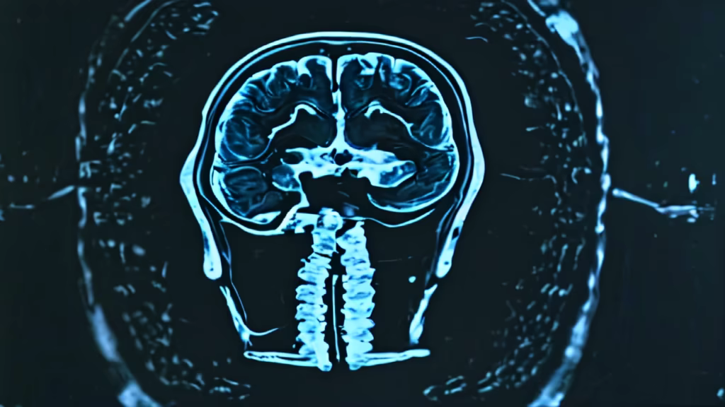
Prompt: THIS IS A COMPUTED TOMOGRAPHY OF BRAIN OF A MIDDLE AGED FEMALE.There is nodular hyperdense heterogeneously enhancing lesion measuring 24x17mm seen in grey white mater junction of left parietal lobe with associated moderate perilesional edema causing effacement of sulcal spaces and, mild effacement of occipital horn. Another similar lesion with mild perilesional edema seen in right inferior cerebellar hemisphere measuring 22x13mm.


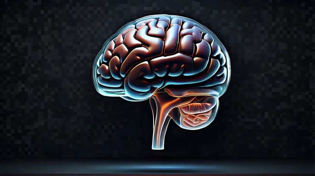
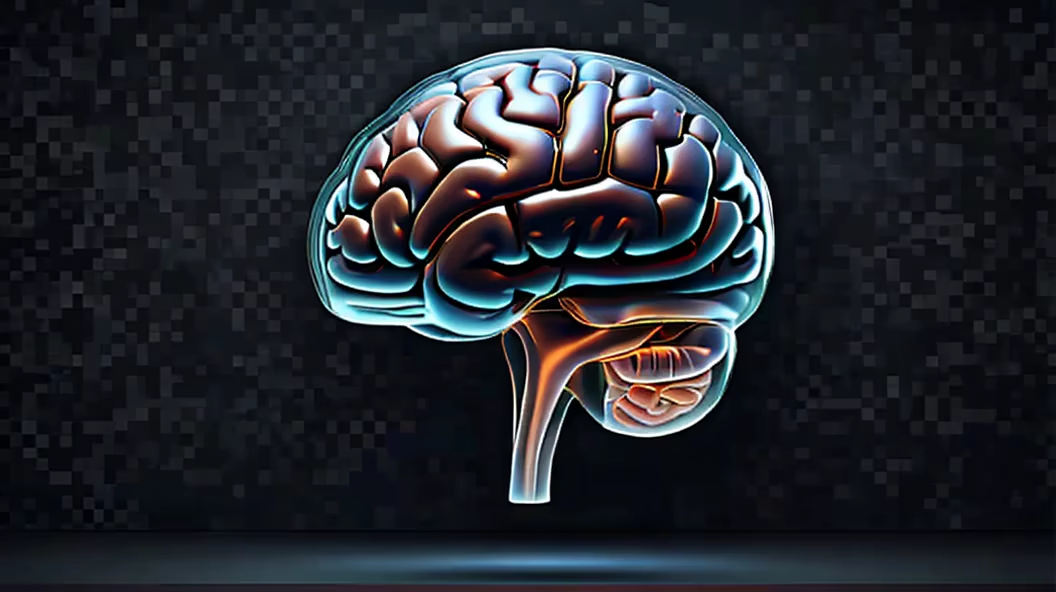
Prompt: A high-fidelity medical x-ray image featuring a sagittal view of a human brain, emphasizing a detailed and accurate portrayal of the brain's convolutions. The x-ray should prominently display a coin embedded in the frontal lobe with utmost clarity. Ensure that the x-ray effect is convincingly strong, completely eliminating any semblance of bony structures in the skull. Pay close attention to the fine details of the brain's convolutions, providing a scientifically accurate representation. Exclude any unnecessary spinal structures from the image. The emphasis should be on achieving a realistic, grayscale x-ray effect that highlights the brain and the embedded coin with precision and clarity.


Prompt: A high-fidelity medical x-ray image featuring a sagittal view of a human brain, emphasizing a detailed and accurate portrayal of the brain's convolutions. The x-ray should prominently display a coin embedded in the frontal lobe with utmost clarity. Ensure that the x-ray effect is convincingly strong, completely eliminating any semblance of bony structures in the skull. Pay close attention to the fine details of the brain's convolutions, providing a scientifically accurate representation. Exclude any unnecessary spinal structures from the image. The emphasis should be on achieving a realistic, grayscale x-ray effect that highlights the brain and the embedded coin with precision and clarity.
Negative: drawing, sketch, painting ,illustration


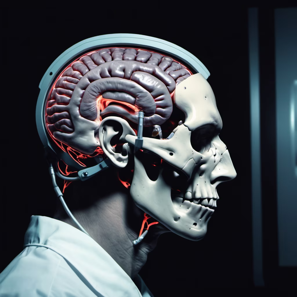
Prompt: A high-fidelity medical x-ray image featuring a sagittal view of a human skull, emphasizing a detailed and accurate portrayal of the brain's convolutions. The x-ray should prominently display a coin embedded in the frontal lobe with utmost clarity. Ensure that the x-ray effect is convincingly strong, completely eliminating any semblance of bony structures in the skull. Pay close attention to the fine details of the brain's convolutions, providing a scientifically accurate representation. Exclude any unnecessary spinal structures from the image. The emphasis should be on achieving a realistic, grayscale x-ray effect that highlights the brain and the embedded coin with precision and clarity.


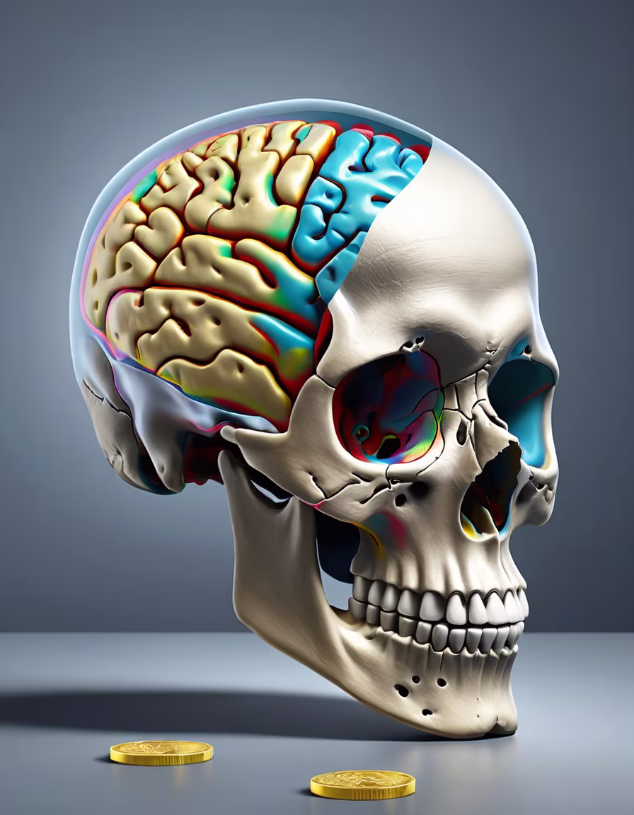
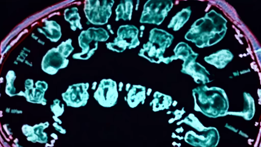
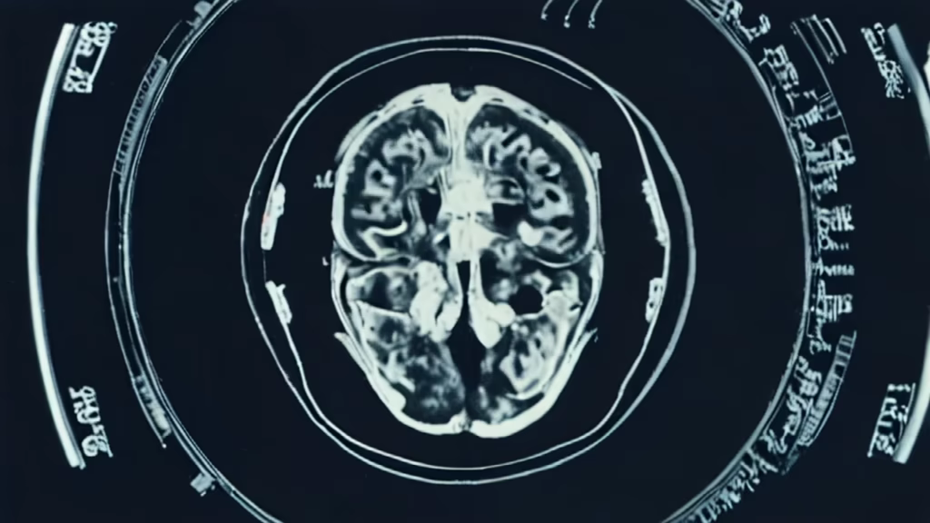
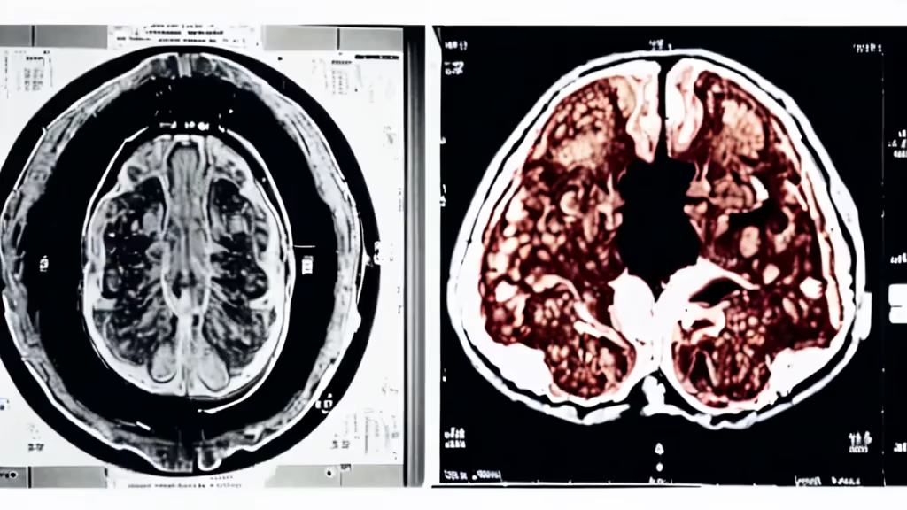
Prompt: Generate a high-resolution medical imaging illustration depicting a CT scan or MRI of a human skull. The image should showcase a clear and realistic x-ray view, emphasizing the presence of a coin embedded in the frontal lobe of the brain. Ensure the coin is seamlessly integrated into the image, with realistic reflections and shading. Pay attention to anatomical accuracy and lighting to create a convincing and visually striking representation.
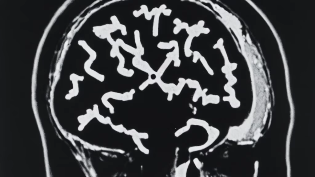


Prompt: Generate a detailed medical x-ray image portraying a sagittal view of a human skull with a distinct focus on the brain. The x-ray should clearly display a coin embedded in the frontal lobe, and the overall image should convey a realistic x-ray effect, with attention to grayscale tones and transparency. Ensure that the x-ray emphasizes the skull's structure, including the outlines of the bones and the intricate details of the brain. Aim for a scientifically accurate and visually compelling representation.


Prompt: The brain should be rendered in intricate detail, with its wrinkled gyri and convoluted sulci highlighted in a vibrant manner. The background should be a vast expanse of space filled with swirling nebulae and radiant stars. Dazzling light should emanate from the brain, illuminating the surrounding space and creating a sense of awe-inspiring energy. which has young asthetic vibe of wise indian monk


Prompt: The brain should be rendered in intricate detail, with its wrinkled gyri and convoluted sulci highlighted in a vibrant manner. The background should be a vast expanse of space filled with swirling nebulae and radiant stars. Dazzling light should emanate from the brain, illuminating the surrounding space and creating a sense of awe-inspiring energ .
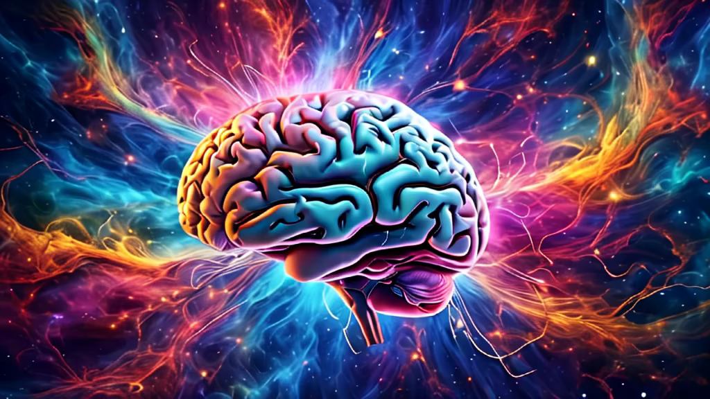
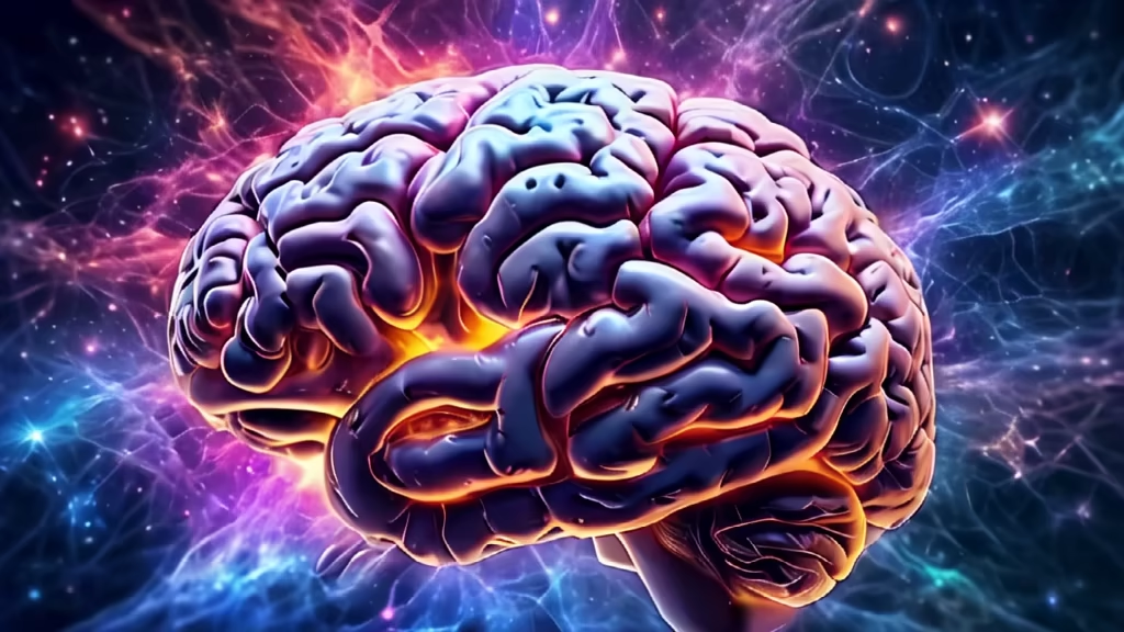


Prompt: The brain should be rendered in intricate detail, with its wrinkled gyri and convoluted sulci highlighted in a vibrant manner. The background should be a vast expanse of space filled with swirling nebulae and radiant stars. Dazzling light should emanate from the brain, illuminating the surrounding space and creating a sense of awe-inspiring energy. which has old asthetic vibe of old wise monk


Prompt: Year:1962. Formal portrait. High detail, Canon EOS DSLR photograph. UHD, 8K resolution, sharp focus, full color, intimate, intricately detailed and extreme close up facial portrait of a fashionable age 29 wife in 1962, showcasing her generously and profusely highlighted blonde hair. She coyly smirks at the camera. Her very thick, softly textured hair fills the viewing screen and is delicately styled in a very large, loose, very tall, very lofty, very highly voluminous bouffant. She has subtle, elegant makeup. Solid, light colored background. Accurate 1962 styling.
Negative: (earrings: 1.6) jewelry, hair beyond camera view, cropped hair, tiara, orange colors, black and white image, nudity, NSFW,
Style: Photographic


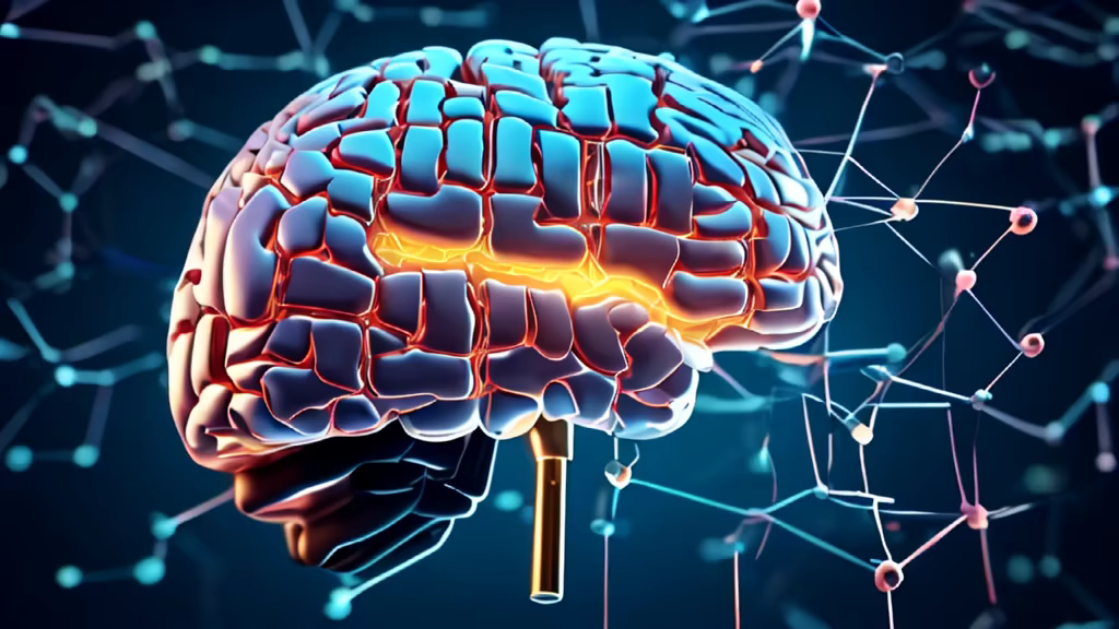
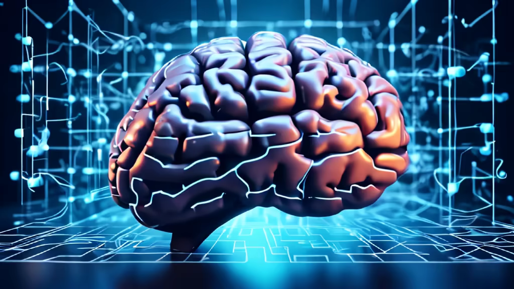
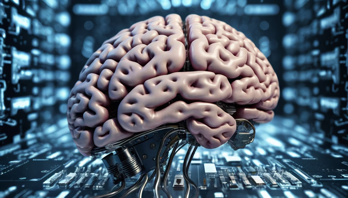
Prompt: I would like an illustration of a brain with a futuristic and technological feel, showcasing the vibrational frequency of brain waves. The brain should appear penetrable, conveying a sense of waves traveling through it, and the brain waves should exhibit a luminous quality, creating a technological vibe.


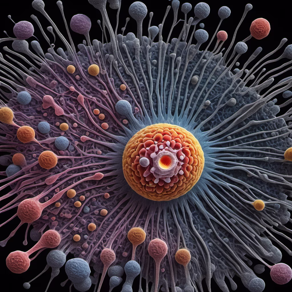
Prompt: A microscopic explorer observed the intricate patterns and designs within populations of bacteria through a high-powered electron microscope, their elaborate cellular structures and organizational motifs expressed through a vivid fusion of micrograph images, abstract artistic renderings and imaginative anthropomorphizations in a thought-provoking multimedia composition illuminated by the cold, clarifying light of scientific inquiry.

Prompt: Generate a high-resolution medical imaging illustration depicting a CT scan or MRI of a human skull. The image should showcase a clear and realistic x-ray view, emphasizing the presence of a coin embedded in the frontal lobe of the brain. Ensure the coin is seamlessly integrated into the image, with realistic reflections and shading. Pay attention to anatomical accuracy and lighting to create a convincing and visually striking representation.
Style: Analog Film


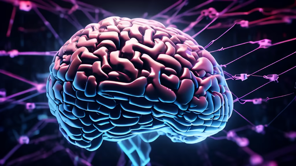
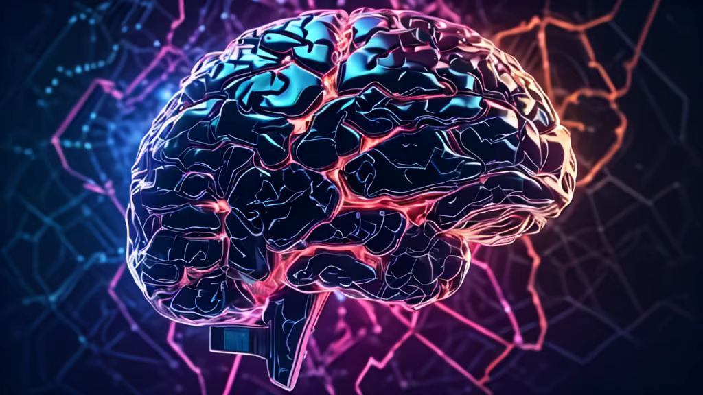
Prompt: Close up of brain, combined, or merging with technology, artificial intelligence, concept image in ultra 16k, ultra enhanced, super sharpen \u0026 detailed image


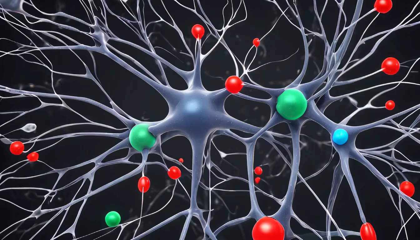
Prompt: Neurons Size: Neurons of different sizes Color: Neurons of different colors Shape: Round, oval, or polygonal shapes Neuronal signals Color: Red, green, or blue colors Shape: Round, oval, or polygonal shapes Direction: Emitted from one neuron, transmitted through synapses to another neuron Synapses Shape: Round or oval shapes Color: Black or gray colors Other Background: Can use white, black, or gray colors Details: Can add additional details, such as the nucleus of the cell on the neuron or the texture on the synapse